Showing posts with label Medicine. Show all posts

Saturday, March 27, 2010

Parkinsonism


Background


Parkinson disease (Parkinson's disease, PD) is a progressive neurodegenerative disorder associated with a loss of dopaminergic nigrostriatal neurons. It is named after James Parkinson, the English physician who described the shaking palsy in 1817.
Parkinson disease is recognized as one of the most common neurological disorders, affecting approximately 1% of individuals older than 60 years. Cardinal features include resting tremor, rigidity, bradykinesia, and postural instability.

Pathophysiology


The major neuropathologic findings in Parkinson disease are a loss of pigmented dopaminergic neurons in the substantia nigra and the presence of Lewy bodies. The loss of dopaminergic neurons occurs most prominently in the ventral lateral substantia nigra. Approximately 60-80% of dopaminergic neurons are lost before the motor signs of Parkinson disease emerge.
Lewy bodies are concentric, eosinophilic, cytoplasmic inclusions with peripheral halos and dense cores. The presence of Lewy bodies within pigmented neurons of the substantia nigra is characteristic, but not pathognomonic, of idiopathic Parkinson disease. Lewy bodies also are found in the cortex, nucleus basalis, locus ceruleus, intermediolateral column of the spinal cord, and other areas. Lewy bodies are not specific to Parkinson disease, as they are found in some cases of atypical parkinsonism, Hallervorden-Spatz disease, and other disorders. Incidental Lewy bodies are found at postmortem in patients without clinical signs of parkinsonism. The prevalence of incidental Lewy bodies increases with age. Incidental Lewy bodies have been hypothesized to represent the presymptomatic phase of Parkinson disease.
No standard criteria exist for the neuropathologic diagnosis of Parkinson disease, as the specificity and sensitivity of the characteristic findings have not been established clearly. Individuals presenting with primary dementia may exhibit neuropathologic features indistinguishable from those of Parkinson disease.
Alpha-synuclein is a major structural component of Lewy bodies. All Lewy bodies stain for alpha-synuclein and most also stain for ubiquitin.


Stages in the development of Parkinson disease-re...

Stages in the development of Parkinson disease-related pathology. Adapted from Braak H, Ghebremedhin E, Rub U, Bratzke H, Del Tredici K. Cell Tissue Res. 2004 Oct;318(1):121-34.


Recent studies demonstrate that Lewy-body pathology in Parkinson disease actually begins in the olfactory bulb and lower brainstem (see image above or 
Media file 4).1 These early stages are associated with premotor symptoms such as loss of sense of smell and rapid eye movement (REM) sleep behavior disorder (RBD).2 The pathology ascends up the brainstem to later involve the midbrain and nigrostriatal dopaminergic neurons. This stage correlates with onset of the motor phase of the disease and patients may exhibit bradykinesia, rigidity, and tremor. The pathology continues to ascend late in the disease to affect the cortex and patients may then exhibit cognitive dysfunction and dementia. 
Motor circuit in Parkinson disease
The basal ganglia motor circuit modulates cortical output necessary for normal movement (see following image or Media file 1).
Schematic representation of the basal ganglia - t...

Schematic representation of the basal ganglia - thalamocortical motor circuit and its neurotransmitters in the normal state. From Vitek J. Stereotaxic surgery and deep brain stimulation for Parkinson's disease and movement disorders. In: Watts RL, Koller WC, eds. Movement Disorders: Neurologic Principles and Practice. New York: McGraw-Hill, 1997:240. Used with kind permission. Copyright, McGraw-Hill Companies, Inc.


Signals from the cerebral cortex are processed through the basal ganglia-thalamocortical motor circuit and return to the same area via a feedback pathway. Output from the motor circuit is directed through the internal segment of the globus pallidus (GPi) and the substantia nigra pars reticulata (SNr). This inhibitory output is directed to the thalamocortical pathway and suppresses movement.

Two pathways exist within the basal ganglia circuit; they are referred to as the direct and indirect pathways. In the direct pathway, outflow from the striatum directly inhibits GPi and SNr. The indirect pathway comprises inhibitory connections between the striatum and the external segment of the globus pallidus (GPe) and the GPe and the subthalamic nucleus (STN). The subthalamic nucleus exerts an excitatory influence on the GPi and SNr. The GPi/SNr sends inhibitory output to the ventral lateral (VL) nucleus of the thalamus. Striatal neurons containing D1 receptors constitute the direct pathway and project to the GPi/SNr. Striatal neurons containing D2 receptors are part of the indirect pathway and project to the GPe.
Dopamine is released from nigrostriatal (SNc) neurons to activate the direct pathway and inhibit the indirect pathway. In Parkinson disease, decreased striatal dopamine causes increased inhibitory output from the GPi/SNr (see following image or Media file 2).
Schematic representation of the basal ganglia - t...

Schematic representation of the basal ganglia - thalamocortical motor circuit and the relative change in neuronal activity in Parkinson disease. From Vitek J. Stereotaxic surgery and deep brain stimulation for Parkinson's disease and movement disorders. In: Watts RL, Koller WC, eds. Movement Disorders: Neurologic Principles and Practice. New York: McGraw-Hill, 1997:241. Used with kind permission. Copyright, McGraw-Hill Companies, Inc.


This increased inhibition of the thalamocortical pathway suppresses movement. Via the direct pathway, decreased striatal dopamine stimulation causes decreased inhibition of the GPi/SNr. Via the indirect pathway, decreased dopamine inhibition causes increased inhibition of the GPe, resulting in disinhibition of the STN. Increased STN output increases GPi/SNr inhibitory output to the thalamus.


Frequency


International


The incidence has been estimated to be 4.5-21 cases per 100,000 population per year. Estimates of Parkinson disease prevalence range from 18-328 per 100,000 population, with most studies yielding a prevalence of approximately 120 per 100,000.

Sex


Parkinson disease is about 1.5 times more common in men than in women.

Age


The incidence and prevalence of Parkinson disease increase with age. The average age of onset is approximately 60 years. Onset in persons younger than 40 years is relatively uncommon.

Clinical


">

History


  • Parkinson disease may have a long premotor phase. Mid-life risk factors for the later development of Parkinson disease include constipation and daytime sleepiness. These may well be the first clinical manifestations of the disease but are nonspecific. Additional features that commonly precede onset of motor signs include decreased sense of smell and REM behavior disorder (RBD).


    • REM behavior disorder is a sleep disorder in which there is a loss of normal atonia during REM sleep.
    • Patients are observed by their bed partners to “act out their dreams” and the partners may note kicking, hitting, talking, or crying out.
    • In one study, 38% of 50-year-old men with REM behavior disorder and no neurologic signs went on to develop Parkinsonism.3
    • REM behavior disorder is common throughout the course of Parkinson disease.
  • Onset of motor signs in Parkinson disease is typically asymmetric, with the most common initial finding being an asymmetric resting tremor in an upper extremity. About 20% of patients first experience clumsiness in one hand. Over time, patients notice symptoms related to progressive bradykinesia, rigidity, and gait difficulty.
  • Tremor usually begins in one upper extremity and initially may be intermittent. As with most tremors, the amplitude increases with stress and resolves during sleep. After several months or years, the tremor may affect the extremities on the other side, but asymmetry is usually maintained. Parkinson disease tremor may also involve the lower extremities, tongue, lips, or chin.
  • The initial symptoms of Parkinson disease may be nonspecific and include fatigue, depression, constipation, and sleep problems.
  • Some patients experience a subtle decrease in dexterity and may notice a lack of coordination with activities such as playing golf or dressing.
  • Some patients complain of aching or tightness in the calf or shoulder region.
  • The first affected arm may not swing fully when walking, and the foot on the same side may scrape the floor.
  • Over time, axial posture becomes progressively flexed and strides become shorter.
  • Decreased swallowing may lead to excess saliva in the mouth and ultimately drooling.
  • Symptoms of autonomic dysfunction are common and include constipation, sweating abnormalities, sexual dysfunction, and seborrheic dermatitis.
  • Sleep disturbances are common.
  • The best clinical predictors of a pathology diagnosis of Parkinson disease are the following:


    • Asymmetry
    • Presence of resting tremor
    • Good response to dopamine replacement therapy
  • Long-term disability in Parkinson disease is usually related to dementia and balance dysfunction.

Physical


The 3 cardinal signs of Parkinson disease are resting tremor, rigidity, and bradykinesia. Of these cardinal features, 2 of 3 are required to make the clinical diagnosis. Postural instability (balance dysfunction) is the fourth cardinal sign, but it emerges late in the disease, usually after 8 years or more.
  • The characteristic Parkinson disease tremor is present and most prominent with the limb at rest.


    • The usual frequency is 3-5 Hz.
    • The tremor may appear as a pill-rolling motion of the hand or a simple oscillation of the hand or arm.
    • The same tremor may be observed with the arms outstretched (position of postural maintenance) and a less prominent, higher frequency kinetic tremor is also common.
  • Rigidity refers to an increase in resistance to passive movement about a joint.


    • The resistance can be either smooth (lead pipe) or oscillating (cogwheeling).
    • Cogwheeling is thought to reflect tremor rather than rigidity and may be present with tremors not associated with an increase in tone (ie, essential tremor).
    • Rigidity usually is tested by flexing and extending the patient's relaxed wrist.
    • Rigidity can be made more obvious with voluntary movement in the contralateral limb.
  • Bradykinesia refers to slowness of movement but also includes a paucity of spontaneous movements and decreased amplitude of movement. Bradykinesia is also expressed as micrographia (small handwriting), hypomimia (decreased facial expression), decreased blink rate, and hypophonia (soft speech).
  • Postural instability refers to imbalance and loss of righting reflexes. Its emergence is an important milestone, because it is poorly amenable to treatment and a common source of disability in late disease.
  • Patients may experience freezing when starting to walk (start-hesitation), during turning, or while crossing a threshold, such as going through a doorway.
  • Dementia generally occurs late in Parkinson disease and affects 15-30% of patients. Short-term memory and visuospatial function may be impaired, but aphasia is not present. Cognitive dysfunction within a year of onset of motor features suggests a diagnosis of Lewy body disease, a disease closely related to Parkinson disease and marked by the presence of cortical Lewy bodies. See Parkinson Disease Dementia for more information.

Causes


Most cases of idiopathic Parkinson disease are believed to be due to a combination of genetic and environmental factors. At both ends of the spectrum are rare cases that appear to be due solely to one or the other.
  • Environmental risk factors associated with the development of Parkinson disease include use of pesticides, living in a rural environment, consumption of well water, exposure to herbicides, and proximity to industrial plants or quarries.
  • Several individuals have been identified who developed parkinsonism after self-injection of 1-methyl-4-phenyl-1,2,3,6-tetrahydropyridine (MPTP).4


    • These patients developed bradykinesia, rigidity, and tremor, which progressed over several weeks and improved with dopamine replacement therapy.
    • MPTP crosses the blood-brain barrier and is oxidized to MPP+ by the enzyme monoamine oxidase (MAO) type B.
    • MPP+ accumulates in mitochondria and interferes with the function of complex I of the respiratory chain.
    • A chemical resemblance between MPTP and some herbicides and pesticides suggested that an MPTP-like environmental toxin might be a cause of Parkinson disease, but no specific agent has been identified. Nonetheless, mitochondrial complex I activity is reduced in Parkinson disease, suggesting a common pathway with MPTP-induced parkinsonism.
  • The oxidation hypothesis suggests that free radical damage, resulting from dopamine's oxidative metabolism, plays a role in the development or progression of Parkinson disease.


    • The oxidative metabolism of dopamine by MAO leads to the formation of hydrogen peroxide. Hydrogen peroxide normally is cleared rapidly by glutathione.
    • If hydrogen peroxide is not cleared adequately, it may lead to the formation of highly reactive hydroxyl radicals that can react with cell membrane lipids to cause lipid peroxidation and cell damage. In Parkinson disease, levels of reduced glutathione are decreased, suggesting a loss of protection against formation of free radicals. Iron is increased in the substantia nigra and may serve as a source of donor electrons, thereby promoting the formation of free radicals.
    • Indices of lipid peroxidation are increased in Parkinson disease.
    • Thus, Parkinson disease is associated with increased dopamine turnover, decreased protective mechanisms (glutathione), increased iron (a pro-oxidation molecule), and evidence of increased lipid peroxidation. This hypothesis raised concern that increased dopamine turnover due to levodopa administration could increase oxidative damage and accelerate loss of dopamine neurons. However, there is no clear evidence that levodopa accelerates disease progression.
  • The role of genetic factors has been studied in twins.


    • If genetic factors are important, concordance in genetically identical monozygotic (MZ) twins will be greater than in dizygotic (DZ) twins, who share only about 50% of genes. Early twin studies generally found low and similar concordance rates for MZ and DZ pairs.
    • In a recent study of 193 twins, overall concordance for MZ and DZ pairs was similar. However, in 16 pairs of twins in whom Parkinson disease was diagnosed at or before age 50 years, all 4 MZ pairs, but only 2 of 12 DZ pairs, were concordant. This suggests that while genetic factors may not be very important when the disease begins after age 50 years, genetic factors appear to be very important when the disease begins at or before age 50 years.
  • The identification of a few large families with apparent familial Parkinson disease sparked further interest in the genetics of the disease.


    • One large family with highly penetrant, autosomal-dominant, autopsy-proven Parkinson disease originated in the town of Contursi in the Salerno province of southern Italy. Of 592 family members, 50 were affected by Parkinson disease. These individuals were characterized by early age of disease onset (mean age 47.5 y), rapid progression (mean age at death 56.1 y), lack of tremor, and good response to levodopa therapy.
    • Linkage analysis incriminated a region in chromosome bands 4q21-23, and sequencing revealed an A-for-G substitution at base 209 of the alpha-synuclein gene. Termed PD-1, this mutation codes for a substitution of threonine for alanine at amino acid 53.
    • Five small Greek kindreds also were found to have the PD-1 mutation. In a German family, a different point mutation in the alpha-synuclein gene (a substitution of C for G at base 88, producing a substitution of proline for alanine at amino acid 30) confirmed that mutations in the alpha-synuclein gene can cause Parkinson disease. A few additional familial mutations in the alpha-synuclein gene have been identified and are now collectively called PARK1. It is now clear that these mutations are an exceedingly rare cause of Parkinson disease.
  • Alpha-synuclein is a major component of Lewy bodies in all Parkinson disease.


    • All Lewy bodies stain for alpha-synuclein, and most also stain for ubiquitin, which conjugates with proteins targeted for proteolysis. Abnormal aggregation of alpha-synuclein into filamentous structures may precede ubiquitization.
    • One hypothesis states that the PD-1 mutation alters the configuration of alpha-synuclein into a ß structure that could aggregate into sheets.
    • All Parkinson disease may be associated with abnormal folding of alpha-synuclein, leading to excessive aggregation and neuronal death.
    • Although sporadic Parkinson disease is not caused by a mutation in the alpha-synuclein gene, active investigation is underway into proteins that interact with alpha-synuclein, including those that guide, promote, or prevent aggregation of the protein.
    • As Parkinson disease, dementia with Lewy bodies, and multiple system atrophy (MSA) all exhibit Lewy bodies that stain for alpha-synuclein, they have been designated "alpha-synucleinopathies."
  • A recent hypothesis suggests that Parkinson disease is caused by abnormalities of the proteosome system, which is responsible for clearing abnormal proteins.
  • Several homozygous deletions in a gene dubbed parkin (PARK2), which is located on chromosome 6, have been found to cause autosomal-recessive juvenile parkinsonism (AR-JP). This form of parkinsonism differs pathologically from Parkinson disease in that no Lewy bodies are found in the substantia nigra.
  • Several other gene abnormalities have been identified in families with Parkinson disease and these are designated PARK3 -PARK12.
  • It has been estimated that all currently known genetic causes of Parkinson disease account for less than 5% of Parkinson disease cases.

Sunday, August 30, 2009

Saturday, August 29, 2009

Origin of swine flu virus


Researchers use evolutionary history to trace the early days of the pandemic.Closely related forms of the H1N1 strain of influenza virus circulated undetected in swine for years, a study published online June 11 in Nature reports. The virus, which has spread to multiple continents, has now been classified by the World Health Organization as a pandemic. "Based on this report, we had a virus circulating in pigs for 10 years and nobody knew anything about it because we were not doing proper surveillance," says Daniel Perez, an influenza expert at the University of Maryland in College Park.

Researchers traced the sordid past of the H1N1 virus by comparing mutations among different strains of the virus. Genetic sequences of 15 swine influenzas from Hong Kong and two human H1N1 viruses were compared with 796 sequences representing a large spectrum of related strains from humans, birds and pigs.

Analyzing numbers of mutations allowed an international team of researchers to estimate how long ago the strains first existed. Virus strains more than 90 percent identical to the current H1N1 strain were circulating in pigs between 9.2 and 17.2 years ago, the researchers found. The current strain "evidently spread without anyone noticing it for 10 years," says Michael Worobey, an evolutionary biologist at the University of Arizona in Tucson and one of the study's authors. "We need to spend more energy looking at what's in pigs."

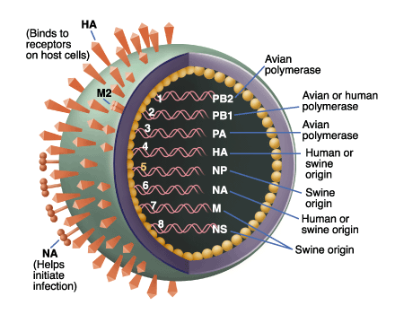
"Any estimate like this has a certain amount of uncertainty to it," Worobey says. Although the numbers are not exact, he says, the data clearly show that a similar version of the virus was around long before anyone was aware of it. The report also shows that each bit of the current virus's DNA had been circulating on its own and primarily in pigs for years before combining to form the virus responsible for the current pandemic. Some genes have been in pigs for decades. "Across the genome, this is something that came from pigs," Worobey says.

Some of these DNA segments came from a North American swine influenza virus, which itself is made of bits of avian, human and swine influenzas (called a triple-reassortant strain). Other segments came from Eurasian swine with avian virus components. The combination of the triple-reassortant strain from North America and the avianlike strain from Eurasia probably happened as live pigs were transported between North America and Eurasia, the authors say.

"We can do all the surveillance we want in humans, but if we really want to prevent pandemic influenza..., a fundamental change in efforts on the animal health side has to be made," Perez says. On the same day the new report appeared, the World Health Organization classified the H1N1 outbreak as a pandemic, defined as showing sustained person-to-person transmission in many parts of the world.

WHO Director-General Margaret Chan said that the organization is raising the alert level after determining that flu cases are now showing up in people who didn't bring it from another region and weren't in contact with such travelers. "Further spread is considered inevitable," Chan said in a news conference.

"This does not mean that there is any difference in the level of severity of the flu," he said. Rather, the pandemic label "is important because it does send the strong message that the virus is here, it's in all likelihood here to stay, and it's important that we continue our aggressive efforts to prepare and respond." So far no decision has been made to mobilize pharmaceutical companies to start mass-producing vaccines aimed specifically at the novel H1N1 virus. But preliminary steps to make that a seamless move have already been taken.

Source: www.sciencenews.org

H1N1 fever- Swine flu Doubts


As the Swine flu is at its kill, everyone is having many doubts. You can clear many of them here

How do you catch H1N1 (swine) flu?

Spread of H1N1 (swine) flu can occur in two ways:

Through contact with infected pigs or environments contaminated with swine flu viruses.
Through contact with a person with H1N1 (swine) flu. Human-to-human spread of H1N1 (swine) flu has been documented also and is thought to occur in the same way as seasonal flu. Influenza is thought to spread mainly person-to-person through coughing or sneezing of infected people.


How does this new H1N1 virus spread?

Spread of this H1N1 virus is thought to be happening in the same way that seasonal flu spreads. Flu viruses are spread mainly from person to person through coughing or sneezing by people with influenza. Sometimes people may become infected by touching something with flu viruses on it and then touching their mouth or nose.


Are there medicines to treat H1N1 (swine) flu?

Yes. CDC recommends the use of oseltamivir (brand name Tamiflu ®) or zanamivir (brand name Relenza ®) for the treatment and/or prevention of infection with these H1N1 (swine) influenza viruses. Antiviral drugs are prescription medicines (pills, liquid or an inhaler) that fight against the flu by keeping flu viruses from reproducing in your body. If you get sick, antiviral drugs can make your illness milder and make you feel better faster. They may also prevent serious flu complications. For treatment, antiviral drugs work best if started soon after getting sick (within 2 days of symptoms).


How long can an infected person spread H1N1 (swine) flu to others?

People with H1N1 (swine) influenza virus infection should be considered potentially contagious as long as they are symptomatic and possible for up to 7 days following illness onset. Children, especially younger children, might potentially be contagious for longer periods


What can I do to protect myself from getting sick?

There is no vaccine available right now to protect against H1N1 (swine) flu. There are everyday actions that can help prevent the spread of germs that cause respiratory illnesses like influenza. Take these everyday steps to protect your health:

Cover your nose and mouth with a tissue when you cough or sneeze. Throw the tissue in the trash after you use it.
Wash your hands often with soap and water, especially after you cough or sneeze. Alcohol-based hand cleaners are also effective.
Avoid touching your eyes, nose or mouth. Germs spread this way.
Try to avoid close contact with sick people.
If you get sick with influenza, CDC recommends that you stay home from work or school and limit contact with others to keep from infecting them.


How can someone with the flu infect someone else?

Infected people may be able to infect others beginning 1 day before symptoms develop and up to 7 or more days after becoming sick. That means that you may be able to pass on the flu to someone else before you know you are sick, as well as while you are sick.


Can people catch H1N1 (swine) flu from eating pork?

No. H1N1 (swine) influenza viruses are not transmitted by food. You can not get H1N1 (swine) influenza from eating pork or pork products. Eating properly handled and cooked pork and pork products is safe. Cooking pork to an internal temperature of 160°F kills the H1N1 (swine) flu virus as it does other bacteria and viruses


Do pigs carry this virus and can I catch this virus from a pig?

At this time, there is no evidence that swine in the United States are infected with this new virus. However, there are flu viruses that commonly cause outbreaks of illness in pigs. Most of the time, these viruses do not infect people, but influenza viruses can spread back and forth between pigs and people.


How long can influenza virus remain viable on objects (such as books and doorknobs)?

Studies have shown that influenza virus can survive on environmental surfaces and can infect a person for up to 2-8 hours after being deposited on the surface.


What surfaces are most likely to be sources of contamination?

Germs can be spread when a person touches something that is contaminated with germs and then touches his or her eyes, nose, or mouth. Droplets from a cough or sneeze of an infected person move through the air. Germs can be spread when a person touches respiratory droplets from another person on a surface like a desk, for example, and then touches their own eyes, mouth or nose before washing their hands.


Is there a risk from drinking water?

Tap water that has been treated by conventional disinfection processes does not likely pose a risk for transmission of influenza viruses. Current drinking water treatment regulations provide a high degree of protection from viruses. No research has been completed on the susceptibility of the novel H1N1 flu virus to conventional drinking water treatment processes. However, recent studies have demonstrated that free chlorine levels typically used in drinking water treatment are adequate to inactivate highly pathogenic H5N1 avian influenza. It is likely that other influenza viruses such as novel H1N1 would also be similarly inactivated by chlorination. To date, there have been no documented human cases of influenza caused by exposure to influenza-contaminated drinking water.


Can the new H1N1 flu virus be spread through water in swimming pools, spas, water parks, interactive fountains, and other treated recreational water venues?

Influenza viruses infect the human upper respiratory tract. There has never been a documented case of influenza virus infection associated with water exposure. Recreational water that has been treated at CDC recommended disinfectant levels does not likely pose a risk for transmission of influenza viruses. No research has been completed on the susceptibility of the H1N1 influenza virus to chlorine and other disinfectants used in swimming pools, spas, water parks, interactive fountains, and other treated recreational venues. However, recent studies have demonstrated that free chlorine levels recommended by CDC (1–3 parts per million [ppm or mg/L] for pools and 2–5 ppm for spas) are adequate to disinfect avian influenza A (H5N1) virus. It is likely that other influenza viruses such as novel H1N1 virus would also be similarly disinfected by chlorine.


Can H1N1 influenza virus be spread at recreational water venues outside of the water?

Yes, recreational water venues are no different than any other group setting. The spread of this novel H1N1 flu is thought to be happening in the same way that seasonal flu spreads. Flu viruses are spread mainly from person to person through coughing or sneezing of people with influenza. Sometimes people may become infected by touching something with flu viruses on it and then touching their mouth or nose.

Monday, August 10, 2009

What is deja vu?


The term deja vu is French and means, literally, "already seen." Those who have experienced the feeling describe it as an overwhelming sense of familiarity with something that shouldn't be familiar at all. Say, for example, you are traveling to England for the first time. You are touring a cathedral, and suddenly it seems as if you have been in that very spot before. Or maybe you are having dinner with a group of friends, discussing some current political topic, and you have the feeling that you've already experienced this very thing -- same friends, same dinner, same topic. Ddeja vu occurs most often in young people.

The phenomenon is rather complex, and there are many different theories as to why deja vu happens. Swiss scholar Arthur Funkhouser suggests that there are several "deja experiences" and asserts that in order to better study the phenomenon, the nuances between the experiences need to be noted. In the examples mentioned above, Funkhouser would describe the first incidence as deja visite ("already visited") and the second as deja vecu ("already experienced or lived through"). As much as 70 percent of the population reports having experienced some form of deja vu. A higher number of incidents occurs in people 15 to 25 years old than in any other age group. Deja vu has been firmly associated with temporal lobe epilepsy. Reportedly, deja vu can occur just prior to a temporal-lobe seizure. People suffering a seizure of this kind can experience deja vu during the actual seizure activity or in the moments between convulsions.

Since deja vu occurs in individuals with and without a medical condition, there is much speculation as to how and why this phenomenon happens. Several psychoanalysts attribute deja vu to simple fantasy or wish fulfillment, while some psychiatrists ascribe it to a mismatching in the brain that causes the brain to mistake the present for the past. Many parapsychologists believe it is related to a past-life experience. Obviously, there is more investigation to be done

Man with tiny brain


A man with an unusually tiny brain manages to live an entirely normal life despite his condition, which was caused by a fluid build-up in his skull. Scans of the 44-year-old man's brain showed that a huge fluid-filled chamber called a ventricle took up most of the room in his skull, leaving little more than a thin sheet of actual brain tissue (see image, right).

"It is hard for me [to say] exactly the percentage of reduction of the brain, since we did not use software to measure its volume. But visually, it is more than a 50% to 75% reduction," says Lionel Feuillet, a neurologist at the Mediterranean University in Marseille, France. Feuillet and his colleagues describe the case of this patient in The Lancet. He is a married father of two children, and works as a civil servant.

Not retarded

The man went to a hospital after he had mild weakness in his left leg. When Feuillet's staff took his medical history, they learned that, as an infant, he had had a shunt inserted into his head to drain away hydrocephalus - water on the brain. The shunt was removed when he was 14. But the researchers decided to check the condition of his brain using computed tomography (CT) scanning technology and another type of scan called magnetic resonance imaging (MRI). They were astonished to see "massive enlargement" of the lateral ventricles - usually tiny chambers that hold the cerebrospinal fluid that cushions the brain. Intelligence tests showed the man had an IQ of 75, below the average score of 100 but not considered mentally retarded or disabled. "The whole brain was reduced - frontal, parietal, temporal and occipital lobes - on both left and right sides. These regions control motion, sensibility, language, vision, audition, and emotional and cognitive functions," Feuillet told New Scientist.

Brain adaptation

The findings reveal "the brain is very plastic and can adapt to some brain damage occurring in the pre- and postnatal period when treated appropriately," he says. "What I find amazing to this day is how the brain can deal with something which you think should not be compatible with life," comments Max Muenke, a paediatric brain defect specialist at the National Human Genome Research Institute in Bethesda, Maryland, US. "If something happens very slowly over quite some time, maybe over decades, the different parts of the brain take up functions that would normally be done by the part that is pushed to the side," adds Muenke, who was not involved in the case.

Monday, August 3, 2009

Rabies - Hydrophobia

Rabies is caused by a bite from an infected animal but occasionally by other forms of contact. In some countries it is a significant killer of livestock. The rabies virus makes its way to the brain by following the peripheral nerves.

RABIES OR hydrophobia is a viral neuroinvasive disease that causes acute encephalitis (inflammation of the brain) in warm-blooded animals. It is zoonotic (ie transmitted by animals), most commonly by a bite from an infected animal but occasionally by other forms of contact. It is fatal if left untreated. In some countries it is a significant killer of livestock.

The rabies virus makes its way to the brain by following the peripheral nerves. The incubation period of the disease depends on how far the virus must travel to reach the central nervous system, usually taking a few months. Once the infection reaches the central nervous system and symptoms begin to show, the untreated infection is usually fatal within days. In the beginning stages of rabies, the symptoms are malaise, headache and fever, while in later stages it includes acute pain, violent movements, uncontrolled excitements, depressions and the inability to swallow water (hence the name hydrophobia).


In the final stages, the patient begins to have periods of mania and lethargy, and coma. Death generally occurs due to respiratory insufficiency. The term is derived from the Latin rabies, "madness." This, in turn, may have come from the Sanskrit rabhas, "to do violence.

The Greeks derived the word "lyssa," which is derived from "lud" or "violent," this terminology is used in the name of the genus of rabies lyssavirus. The rabies virus is the type species of the Lyssavirus genus, which encompasses other similar viruses.

Lyssa-viruses have helical symmetry, with a length of about 180nm and a cross-sectional diameter of about 75nm. These viruses are enveloped and have a single stranded RNA genome with negative-sense. The genetic information is packaged as a ribonucleoprotein complex, in which RNA is tightly bound by the viral nucleoprotein.

The RNA genome of the virus encodes five genes whose order is highly conserved. These genes are nucleoprotein (N), phosphoprotein (P), matrix protein (M), glycoprotein (G) and the viral RNA polymerase (L). From the point of entry, the virus travels quickly along the neural pathways into the central nervous system (CNS) and then further into other organs. The salivary glands receive high concentrations of the virus thus allowing further transmission.